


Surgeon Performed Ultrasound
Does the breast surgeon offer surgeon performed office ultrasound ?
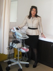
Jane O’Brien is proud to be one of the few breast services to hold a certificate of accreditation for breast ultrasound, (having fulfilled the requirements of the Diagnostic Imaging Accreditation Scheme Standards) and is therefore able to offer patients the additional service of surgeon performed ultrasound using a mobile, portable ultrasound machine. Used as an extension of the clinical examination it increases diagnostic accuracy and most importantly for the patient helps to streamline the process during what is an anxious time. Breast ultrasound is rapidly becoming an indispensable component of the surgeon’s armamentarium for the diagnosis and treatment of breast disease. It is not a substitute for a formal radiologist performed ultrasound; it is complementary, enabling the surgeon to correlate the clinical examination with the ultrasound findings by palpating the lump or clinical region of concern while scanning.
 Ultrasound is an imaging modality which uses high frequency sound waves to scan the breast tissue. The vibrations from these sound waves are reflected off the breast tissue and transformed into electrical signals that show up as an image on a screen. As a diagnostic tool, ultrasound is often used in conjunction with conventional mammography. The ultrasound itself does not use radiation. New advances in ultrasound technology have led to the development of high performance, mobile ultrasound devices which enable breast surgeons to use a hand held ultrasound in the consulting room during breast examinations. The breast surgeon using office ultrasound does have the advantage of viewing ‘dynamic’ images which is preferable to the viewing of ‘static’ images as often done by radiologists. The portable ultrasound becomes an extension of the physical examination of the breast-an extension of the surgeon’s hand or the “stethoscope of the breastsurgeon”.
In almost every area of surgery specialty, ultrasound is being increasingly utilized and incorporated into daily clinical practice by surgeons. This has been facilitated by advances in ultrasound technology which have seen the development of many high quality small portable ultrasound units, which are extremely user friendly. Office ultrasound is particularly applicable and relevant for the breast surgeon who is able to use this technology to enhance the assessment of and streamline the management of breast patients. Breast ultrasound should be considered an extension of the physical examination and can often be used to provide immediate reassurance for symptomatic women. Office ultrasound assists the breast surgeon to undertake fine needle aspiration biopsies and core biopsies. It also enables the better pre-operative assessment of breast cancer patients and it may help in decision-making regarding optimal surgical management. It can also be a useful tool in the management of breast abscesses through repeated aspirations.
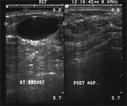
Simple cyst of the right breast before and after needle aspiration by ultrasound guidance.
One of the most useful applications of ultrasound scanning of the breast is in differentiating between innocent cysts (fluid filled lumps) and more sinister solid lumps (potential cancers). When a woman visits a breast surgeon with a lump in the breast this is a very anxious time for her, and being able to immediately after the physical examination of the breast, perform an on the spot ultrasound and confirm that a lump is indeed a simple cyst is extremely useful. The ultrasound can then if necessary be used to guide needle placement for aspiration of the fluid within the cyst with immediate resolution of the lump and the patient can leave the initial consultation with the reassurance that the lump was not only innocent in nature, but has been successfully dealt with.
The mobility and portability of the ultrasound machine make it also ideal for use also by the breast surgeon in the operating theatre, where it helps to identify and delineate the extent of breast lesions prior to removal, allowing on –table needle localisation and can also be used to examine the specimen once removed to ensure adequate margins. It is a non invasive test that is rapid and easily repeatable. The images can be stored on the hard drive of the ultrasound machine or hard copies can be produced using the attached printer for storage in the patient’s records.
It is important that breast surgeons incorporate ultrasound into their practice to remain relevant to the management of women with breast disease. Breast surgeons using ultrasound will remain at the forefront of newer diagnostic and therapeutic interventions and Jane O’Brien is very pleased to be fully accredited to offer patients this service.
Error: Contact form not found.